Leucocytes divide into main two groups:
- Granulocytes - (neutrophils, basophils, eosinophils)
- Agranulocytes - (lymphocytes, monocytes)
Granulocytes have granules in their cytoplasm that have functional importance. However, some recent studies also suggest that lymphocytes and monocytes also have granules.
Products of immune cells have a role in mediating immunity against many pathogens like bacteria, viruses, parasites, allergens, tumor cells, etc.
1. Neutrophils
Neutrophils are abundantly present in blood as compared to other leucocytes and motile serves as the first line of defense.
Neutrophils are produced from bone marrow pluripotent stem cells(myeloid progenitors) and bone marrow produces more neutrophils when needed. (IL-3 help to grow and differentiate pluripotent stem cell to various immune cells)
Neutrophils have a short life span after getting entry into the blood.
Neutrophils are circulating in the blood and when the inflammatory process is going on in certain tissue then it migrates to tissue and helps to eliminate pathogens.
Chemotactic factors can be chemotactic neutrophils. IL-8 has a chemotactic activity that is released from the inflammatory area.
Neutrophils have phagocytic activity through different pathways. ( oxygen dependant, oxygen-independent, various enzymes-lysozymes, peptidases, collagenases, lactoferrin, etc.
After digesting the component of the pathogen, neutrophils are released into the surrounding tissue. But macrophages present into MHC-2 molecule and present to Th(T-helper) cells.
The normal value of neutrophils in blood
- Dog/cat :- 60-77%
- Horse:- 52-70%
- Cattle/buffalo:- 25-35%
| Electron microscopic image of neutrophils |
2. Eosinophils
Eosinophils also contain granules in their cytoplasm. It has phagocytic activity but is not as much important as neutrophils.
It has a very important role in parasitic infection. It damages the membrane of parasites.
ECF(Eosinophils chemotactic factor increase the number of eosinophils at the site of infection)
In the bone marrow, IL-5 induces differentiation of progenitor cells to eosinophils.
IgE antibody is for from B cell under influences of IL-4 by class switching.
Eosinophils have Fc p[ortion of IgE antibody and it coated to the parasite. Damaging parasite with ADCC(antibody dependant cell cytotoxicity) pathway.
Eosinophils trigger when it coated with IgE and release basic protein which is toxic to helminth.
The normal value of eosinophils in blood
- Dog/cat :- 2-10%
- Horse:- 0-7%
- Cattle/buffalo:- 2-20%
 |
| Electron microscopic image of eosinophils |
3. Basophils
Basophil progenitor cells differentiate and form basophil and release in blood. Basophil and its advanced structure both are involved in an allergic response.
Mast cell precursors are released into blood and undifferentiated until they leave the blood.
Mast cells are present in the skin, connective tissue, epithelial layer of the respiratory, urogenital, and digestive tract.
The most active product of mast cells is histamine and very important role in allergy. Histamine bind to endothelial cells, stomach cell, autocrine manner, and work also as neurotransmission by this way showing its effect.
Mainly 4 types of histamine receptor present
- H1 receptor:- Involve in allergic inflammation
- H2 receptor:- Gastric secretion
- H3 receptor:- Neurotransmission
- H4 receptor:- Immunomodulation
The normal value of basophil in the blood
- Dog/cat :- 0-1%
- Horse:- 0-12%
- Cattle/buffalo:- 0-2%
 |
| Electron microscopic image of basophils |
4. Lymphocytes
In lymphocytes, 3 cells are there - T cells, B cells, NK(natural killer) cells.
T cells, B cells are involved in adaptive immunity while NK cells are involved in innate immunity.
Specific immunity is due to its memory cells development after activation of such Cells. TCR and BCR are part of the specificity of T cell and B cell respectively.
Lymphocytes develop in bone marrow from lymphoid progenitor cells.
T cells mature in the thymus while B cells mature in bone marrow in mammals but in birds, they mature in the bursa of Fabricius.
Mature in context to get specificity to antigen CD molecule development. Self-antigen-specific cells are destroyed in the primary lymphoid organ.
Two types of T cells
- Cytotoxic t cell
- T helper cell
Cytotoxic T(Tc cell) cells only activate when TCR recognizes antigen and are co-stimulated by CD-8 molecule which recognizes MHC-1 molecule.
Activated Tc cells secrete perforins and granzymes which kill the cell.
While helper T cell only activates when TCR recognizes antigen and is co-stimulated by Cd-4 molecule which recognizes MHC-2 molecule.
T helper(Th cell) cell is two type
- Th1 cell
- Th2 cell
Th1 cells participate in cell-mediated immunity while Th2 cells involve in humoral immunity.
Macrophages secrete IL-12 and Th naive cells differentiate into Th1 cells instead of Th2 cells. Th1 cells secrete IFN-gamma which is a potent macrophage activator.
Th2 cells involve in humoral response by secreting IL-4 and IL-5. This will activate and differentiate B cells and produce antibody-secreting plasma cells.
B cells have BCR on their surface and the receptor is IgD or IgM antibody. Which have specific specificity to antigen and activate when antigen bind in it.
B cell and T cell have diversity more than C individual.
B cells activate and differentiate into antibody-secreting plasma cells. Antibody switching is based on stimulants by certain chemical compounds.
Antibodies then participate in humoral response and work as an opsonin and facilitate phagocytosis.
NK cells have a very important role in immunity against cancer cells.
The normal value of Lymphocytes in blood
- Dog/cat :- 12-30%
- Horse:- 21-42%
- Cattle/buffalo:- 45-75%
 |
| T cell electron microscopic image |
| NK cell electron microscopic image |
5. Monocytes
Monocytes develop from granulocytes monocyte progenitor cells.
Monocytes circulate in the blood and it differentiates into macrophage which is present in the tissue. Two types of free macrophage circulate in the blood and the other type of macrophage is in certain tissue and stays.
A macrophage of tissue:-
- Kuffer cells - Liver
- Alveolar macrophage - Lung
- Histiocytes - Connective tissue
- Mesangial cells - Kidney
- Osteoclast - Bone
- Microglial cells - Brain
Macrophage phagocyte the pathogen and form phagosome which degrades pathogen to many molecules by enzymes and oxygen dependant and independent pathway.
Macrophages then present into their surface molecule(MHC-2). This will activate the Th cell.
When macrophage engulfs certain bacteria like mycobacterium, it secretes IL-12 which differentiate Th naive cell to Th1 cell.
Simple bacterial infections trigger Th2 cells and facilitate humoral response. In which IL-12 is not secreted and Th2 cells secrete IL-4 & IL-5 instead of IFN-gamma.
The normal value of monocytes in blood
- Dog/cat :- 2-4%
- Horse:- 0-7%
- Cattle/buffalo:- 2-7%
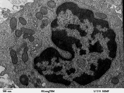 |
| Electron microscopic image of Macrophage |
Other cells involve in immunity is dendritic cells which are potent antigen-presenting cells and possess MHC-2 molecule on their membrane.
Dendritic cells are also formed from myeloid as well as lymphoid progenitor cells.
